Keyhole Surgeries
Laparoscopy and Laparoscopic Surgeries

Laparoscopic surgery , also called minimally invasive surgery (MIS), bandaid surgery, keyhole surgery is a modern surgical technique in which operations in the abdomen are performed through small incisions (usually 0.5–1.5 cm) as compared to larger incisions needed in traditional surgical procedures.
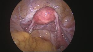
Keyhole surgery uses images displayed on TV monitors for magnification of the surgical elements. There is a direct visualization of the peritoneal cavity, ovaries, outside of the tubes and uterus by using an instrument somewhat like a miniature telescope with a fiber optic system which brings light into the abdomen. A laparoscopy can lead to the diagnosis of many problems which cause infertility including damaged tubes, endometriosis, adhesions and tuberculosis.
A diagnostic laparoscopy, in some clinics is a routine part of the workup in infertile women, in order to complete their evaluation. Generally, the procedure is performed after the basic infertility tests are done, since it is a surgical (invasive) procedure.
Most doctors, these days, try to perform the diagnostic laparoscopy during the post-menstrual phase, when the uterine lining is thin, so that they can combine it with a hysteroscopy at the same time.
The patient is advised not to eat or drink anything for approx. 8-10 hours before the operation. Some tests may also be done before the procedure, to ensure safety for anaesthesia. The keyhole surgery is usually done on a day-care basis. Laparoscopy is done under general anaesthesia so that the patient remains asleep during surgery and does not feel any discomfort.
Bowel Preparation: You may be given instructions regarding this during your preoperative office visit. Bowel preparation is usually recommended for patients with endometriosis, pelvic adhesions or pelvic pain. Preparing the bowel with a purging agent is often followed by an oral antibiotic and enemas. While unpleasant, this procedure minimizes the risk of surgical complications from bowel injury during your surgery.
Patient must shower or bathe the night prior to surgery.
Vaginal Prep: None is usually required.
Nail polish, make-up and jewelry should be removed the night before keyhole surgery.
Wear loose-fitting clothes to prevent any unnecessary pressure on the umbilicus on the day of surgery.
The laparoscopic procedure: First the abdomen is cleansed and draped for the procedure. Then an instrument may be placed in the uterus through the vagina. A gas, such as carbon dioxide or nitrous oxide or air is then allowed to flow into the abdomen just below the belly button. This gas creates a space inside by pushing the abdominal wall and the bowel away from the organs in the pelvic area and makes it easier to see the reproductive organs clearly.
The laparoscope, which is a slender tube, like a miniature telescope, is then inserted through a small incision just below the navel. During the laparoscopy a small probe is placed through another incision in order to move the pelvic organs into clear view. A diagnostic laparoscopy is incomplete without a “second puncture” because, without this second probe, it is not possible to visualize all the structures completely.
During the laparoscopy the entire pelvis is carefully scanned and the organs inspected systematically – the uterus; the ovaries; and the lining of the abdomen, called the peritoneum. In addition to looking for diseases affecting these structures, the doctor also looks for adhesions (bands of scar tissue), endometriosis and tubercles. In case abnormalities are found, the doctor can either try to correct them (operative laparoscopy), or take out bits of tissue for histologic examination (biopsy) with a biopsy forceps. A blue dye (methylene blue) is then injected through the uterus and fallopian tubes to check whether the tubes are open. When the keyhole surgery is complete, the gas is removed and one or two stitches inserted to close the incisions. Since the incisions are so small, often stitches are not needed and they can be closed with Band-Aids.
A diagnostic hysteroscopy is also done at the same time to ensure that the uterine cavity is normal. Most doctors today use videolaparoscopy, in which a video camera is connected to the laparoscope, so that what the surgeon sees can be displayed on a TV monitor. This kind of laparoscopy can be very useful for documentation and record-keeping as well as for patient education.
During operative laparoscopy, many problems which cause infertility can be safely treated through the laparoscope at the same time that the diagnosis is made. When performing operative laparoscopy, additional instruments such as probes, scissors, biopsy forceps, coagulators and suture materials are placed into the abdomen, either through the laparoscope or through two or three additional incisions called “suprapubic punctures”, which are made above the pubis.
Some of the disorders that can be corrected with the help of the procedures above include: releasing scar tissue and / or adhesions from around the fallopian tubes and ovaries; opening blocked tubes; and removing ovarian cysts. Endometriosis can also be destroyed by burning it from the back of the uterus, ovaries, or peritoneum during operative laparoscopy. Under certain circumstances, small fibroid tumors can be removed and ectopic pregnancies can be treated.
After surgery, you will wake up in the recovery room. The nurse will check your blood pressure, pulse and temperature frequently. If you are cold, ask for an extra blanket. The nurse or physician will tell you when you will be allowed to drink something.
Your physician will discuss the findings with your family immediately after the surgical procedure is complete.
Medication will be available for pain or nausea. Pain medication is usually allowed every 3-4 hours. Medication for nausea is usually allowed every 4-6 hours.
Sore Throat: You may experience a sore throat. This is caused by irritation from a tube placed in your throat (trachea) during anaesthesia. It usually lasts for just a few days and can sometimes be helped by throat lozenges.
You will remain in the Recovery Room for approximately three or four hours after the procedure. After you are able to empty your bladder, you will be allowed to go home. You will be given prescriptions to take with you.
The risk of laparoscopy are minimal. But certain conditions increase the possibility of complications. If there has been previous surgery in the abdomen, especially involving the bowel, there is an increased risk. Other conditions that lead to a higher risk of complications are evidence of an infection in the abdomen, a large growth or tumor within the abdomen, and obesity.
Complications among young, healthy women undergoing laparoscopy are rare and occur only in about three out of 1000 cases. These complications can include injuries to structures in the abdomen such as the bowel, a blood vessel or the bladder. Most often, these injuries occur when the laparoscope is placed through the navel. If such an injury occurs during the procedure, the physician can perform major surgery and correct the damage through a longer abdominal incision. Sometimes, complications may arise after surgery. If bleeding or pain appears excessive or if high fever develops, the doctor should be informed.
For any further queries regards the laparoscopic keyhole surgeries, please feel free to contact us at [email protected]
Hysteroscopy and Hysteroscopic Surgeries

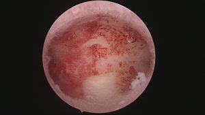
Hysteroscopy is the inspection of the uterine cavity by endoscopy with access through the cervix. It allows for the diagnosis of intrauterine pathology and serves as a method for surgical intervention (operative hysteroscopy). The procedure assists in the diagnosis of various uterine conditions which cause infertility, such as:
- Submucous (internal) fibroids
- Scarring (adhesions or synechiae)
- Endometrial polyps
- Uterine septa and other congenital malformations
Diagnostic hysteroscopy is usually conducted on a day-care basis with either general or local anaesthesia and takes about thirty minutes to perform.
Hysteroscopy has been done in the hospital, surgical centers and the office. It is best done when the endometrium is relatively thin, that is after a menstruation. The first step of hysteroscopy involves cervical dilatation – stretching and opening the canal of the cervix with a series of dilators. Once the dilatation of the cervix is complete, the hysteroscope, a narrow lighted telescope, is passed through the cervix and into the lower end of the uterus. A clear solution (Hyskon or glycine) or carbon dioxide gas is then injected into the uterus through the instrument. This solution or gas expands the uterine cavity, clears blood and mucus away, and enables the surgeon to directly view the internal structure of the uterus.
The doctor systematically examines the lining of the cervical canal; the lining of the uterine cavity; and looks for the internal openings of the fallopian tubes where they enter the uterine cavity – the tubal ostia. A curettage (a surgical scraping of the inside of the uterine cavity) is performed after the hysteroscopy and the endometrial tissue is sent for pathologic examination.
The technique of hysteroscopy has also been expanded to include operative hysteroscopy. Operative hysteroscopy can treat many of the abnormalities found during diagnostic hysteroscopy at the time of diagnosis. The procedure is very similar to diagnostic hysteroscopy except that operating instruments such as scissors, biopsy forceps, electocautery instruments, and graspers can be placed into the uterine cavity through a channel in the operative hysteroscope. Fibroid tumors, scar tissue (synechiae or adhesions), and polyps can be removed from inside the uterus. Congenital abnormalities, such as a uterine septum, may also be corrected through the hysteroscope.
The overall complication rate for diagnostic and operative hysteroscopy is 2% with serious complications occurring in less than 1% of cases. Complications occur rarely during hysteroscopy. In a few cases, infection of the uterus or fallopian tubes can result. Occasionally, a hole may be made through the back of the uterus – a perforation. However, this is usually not a serious problem because the perforation closes on its own. Frequently, when extensive operative hysteroscopy is planned, diagnostic laparoscopy is performed at the same time to allow the surgeon to see the outside as well as the inside of the uterus to try to reduce the risk of accidental uterine perforation. Other possible complications include allergic reactions and bleeding.
Polyps Endometrial or uterine polyps are soft, fingerlike growths which develop in the lining of the uterus (the endometrium). They develop because of excessive multiplication of the endometrial cells, and are hormonally dependent, so that they increase in size depending upon the estrogen level. They can usually be detected on an ultrasound scan if this is done mid-cycle, when estrogen levels are maximal, but are easily missed if the scan is not done at the right time of the menstrual cycle. Polyps are an uncommon but important cause of infertility, because they can easily be removed during hysteroscopic surgery.
Copyright © 2025, Corion. All Rights Reserved.
Website is designed & developed by Phi Brands


